 NA-MIC Project Weeks
Back to Projects List
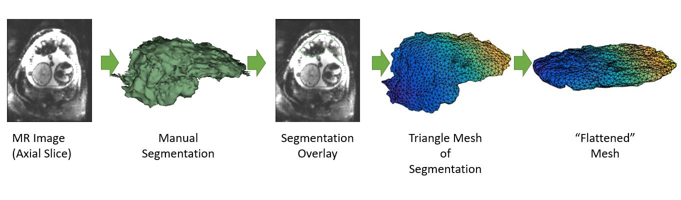
Mapping and Visualizing a Segmented Placenta to a Canonical Template
Key Investigators
- Mazdak Abulnaga (MIT)
- Danielle Pace (MIT)
- Steve Pieper (Isomics)
- Justin Solomon (MIT)
- Polina Golland (MIT)
Project Description
The project seeks to flattened images of the placenta for visualization. Segemented placenta are mapped to a canonical template such as an ellipsoid for visualization of anatomy and function. We aim to build a module in Slicer to establish correspondence between the original volume and the flattened one.
Objective
-
- Map the segmented placenta with minimal distortion.
-
- Map the grayscale intensity from the original volume to the flattened volume.
-
- Build the visualization module in Slicer. Objective is to be able to click on a location in the original volume and automatically move the cursor on the flattened volume.
Approach and Plan
Progress and Next Steps
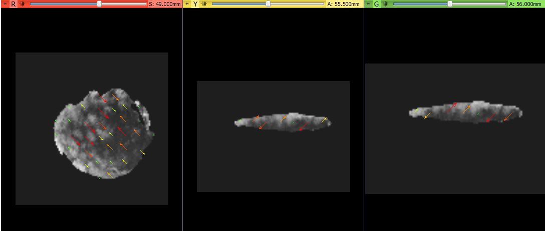
- Created a few test transformations and generated the appropriate displacement fields.
- Saved the displacement fields as NRRD files and successfully loaded them into Slicer.
- Identified bugs in the transformation, it seems there is an inherent rotation that Slicer is unaware of so the field does not apply correctly.
Next Steps:
- Rotation on simple test cases
- Rotation on transformed volumes
- Develop visualization module
Illustrations